

Artlabeling Activity Areolar Connective Tissue a Model Connective Tissue
Introduction
Connective tissue, equally the name implies, is a term given to several different tissues of the torso that serve to connect, support and assistance bind other tissues in the body. Connective tissue can further be cleaved down into three categories: loose connective tissue, dense connective tissue, and specialized connective tissue. Loose connective tissue works to hold organs in place and is made upwards of extracellular matrix and collagenous, elastic and reticular fibers. Dense connective tissue is what makes upward tendons and ligaments and consist of a higher density of collagen fibers. Examples of specialized connective tissues are adipose tissue, cartilage, os, blood, and lymph.
Construction and Function
Loose and dense connective tissue are fabricated up of the following iii fibers: collagen fibers, reticular fibers, and elastin fibers.
Collagen fibers are made up of closely packed sparse collagen fibrils that run a wavy grade in tissues. These parallel fibrils are bundles with flexible proteoglycans to offer an essential mechanical property. They offer flexible only powerful resistance to pulling strength. Specifically, in loose connective tissue, collagen runs in a parallel grade and and so joins to course a larger packet. They split up from each other and join dorsum together at varying locations creating a three-dimensional meshwork. Dense connective tissue such every bit ligaments and tendons are compromised mainly of densely packed collagen fibers.[1]
Reticular fibers, too called argyrophilic fibers, have a restricted abundance in the human torso. They are primarily present in basement epithelial tissue, adipose cells, Schwann and muscle cells, lymphoid tissue and endothelium of hepatic sinusoids. Under microscopy, these reticular fibers are fine, nighttime fibrils that are continuous with the college fibers described in a higher place. The arrangement of these fibers forms a network that underlies the basal lamina layer. At that place is a firm attachment of these fibers to the basal lamina that indicates that, along with the collagen fibers, these fibers create a functional and structural unit that serves to back up tissues. The loose arrangement of these fibers besides provides infinite for molecular motility within the extracellular fluid.[ane]
The last component to discuss is elastin fibers. These fibers have the characteristic holding of elastic recoil. Typically, in loose connective tissue, elastin is a loose network. Their organization and distribution depend on the type of tissue. Concentric elastin fibers are present in the vascular wall to help maintain uniform claret force per unit area. Fibers are as well nowadays in distensible and contractible organs such as the lungs and urinary bladder.[i]
Embryology
Connective tissue arises from the somatic mesoderm. Inductive signals from nearby sclerotome and myotome crusade an upregulation expression of a key transcription factor in tenogenic and ligamentogenic differentiation chosen scleraxis. Several fibroblast factors every bit well equally transforming growth gene-beta accept interest in regulating tendon development. Tendon progenitor cells begin to lay down collagenous fibrils, and these fibrils grow in different directions and beginning to form the tendon fascicle. Tendon fibroblasts reside betwixt collagen fibers. A connective tissue layer called the epitenon surrounds these tendon fascicles to form the complete tendon tissue.[two]
Claret Supply and Lymphatics
Different types of connective tissue have a variable blood supply. Tendons and ligaments, in item, appear partially avascular. They are compromised mainly of densely packed collagen fibers which undergo no metabolic activity and don't require a blood supply. In that location are living cells hidden inside these collagen fibers which require claret supply; however, their book is minimal as compared to the tendons as a whole.[3]
Nerves
All peripheral nervus fibers consist of iii connective tissue layers which serve every bit a protective connective sheath. Epineurium is the outer about layer of dense connective tissue that encloses the unabridged peripheral nervus. Within the epineurium, there are several nerve fascicles which are individually enclosed by the perineum. These fascicles are made upward of myelinated individual nerve fibers that are surrounded by endoneurium.[iv]
Muscles
Individual muscles cells are grouped together to form a fiber. These fibers are further bundled together to form a fascicle, and several of these fascicles become further grouped together to create the entire musculus. Connective tissue exists betwixt every muscle cell, cobweb, and fascicle. At a molecular level, each muscle cell is connected to other musculus cells past a collagenous basement membrane called an endomysium. The fascicles are surrounded past perimysium which further connects to the epimysium which encompasses the entire skeletal musculus and is continuous with the tendon. The collagenous network offset at the level of the endomysium is continuous with the perimysium and the tendon, which allows for an effective and powerful muscle contraction.[five]
Clinical Significance
The following are a pair of examples of clinically significant connective tissue conditions:
Mixed Connective Tissue Disease
Mixed connective tissue affliction (MCTD) is an autoimmune connective tissue disorder characterized by an autoantibody to ribonucleoprotein (RNP). It presents clinically as SLE, systemic sclerosis, and polymyositis. Diagnostic criteria are based on anti-RNP serology with myositis or synovitis plus two of the following: edema of hands, Raynaud's miracle, sclerodactyly/acrosclerosis. Pulmonary symptoms are prevalent in patients with MCTD. Patients may mutter of cough, dyspnea or pleuritic chest hurting. Pulmonary hypertension is the most severe pulmonary consequence and often leads to premature expiry.[6]
Rotator Cuff Injury
The rotator gage is comprised of four tendons localized in the shoulder region. These tendons originate from the following muscles: subscapularis, supraspinatus, infraspinatus, and teres small. Rotator cuff injuries tin nowadays as debilitating pain, reduced shoulder move and role, and shoulder weakness. Treatment is started initially with physical therapy and corticosteroid injections. Surgical techniques are also available for patients that have failed conservative therapy; still, research has demonstrated an equivocal benefit with the surgical arroyo. Patients with rotator cuff tendon injuries are at increased chance for repeated tears through their lifetime.[2]
Review Questions
Figure
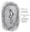
The Ureters, Transverse section of ureter, Fibrous tissue, Longitudinal muscular fibers, Round muscular fibers, Subepithelial connective tissue, Transitional epithelium. Contributed by Gray'south Anatomy Plates

Figure
Connective tissue, dense, adipose, areolar, compact bone, claret. Illustration by Emma Gregory
References
- 1.
-
Ushiki T. Collagen fibers, reticular fibers and elastic fibers. A comprehensive agreement from a morphological viewpoint. Arch Histol Cytol. 2002 Jun;65(two):109-26. [PubMed: 12164335]
- 2.
-
Yang G, Rothrauff BB, Tuan RS. Tendon and ligament regeneration and repair: clinical relevance and developmental image. Nativity Defects Res C Embryo Today. 2013 Sep;99(iii):203-222. [PMC free article: PMC4041869] [PubMed: 24078497]
- three.
-
EDWARDS DA. The blood supply and lymphatic drainage of tendons. J Anat. 1946 Jul;80:147-52. [PMC gratuitous commodity: PMC1272723] [PubMed: 20996686]
- 4.
-
Liu Q, Wang Ten, Yi South. Pathophysiological Changes of Physical Barriers of Peripheral Fretfulness After Injury. Forepart Neurosci. 2018;12:597. [PMC costless article: PMC6119778] [PubMed: 30210280]
- five.
-
Calorie-free N, Champion AE. Characterization of muscle epimysium, perimysium and endomysium collagens. Biochem J. 1984 May 01;219(3):1017-26. [PMC free article: PMC1153576] [PubMed: 6743238]
- six.
-
Pepmueller PH. Undifferentiated Connective Tissue Affliction, Mixed Connective Tissue Disease, and Overlap Syndromes in Rheumatology. Mo Med. 2016 Mar-Apr;113(2):136-40. [PMC costless article: PMC6139943] [PubMed: 27311225]
Source: https://www.ncbi.nlm.nih.gov/books/NBK538534/
0 Response to "Artlabeling Activity Areolar Connective Tissue a Model Connective Tissue"
Post a Comment